Posts

Which foods are good for brain?

Image
  The brain is the most important organ of our body's CNS (Central Nervous System).  It is the hard disk of our central nervous system.  Our brain not only stores memories, but also control our heartbeat, lungs breathing, kidney filtration, and also allows us to think, move and feel.  As the brain is the main organ of the nervous system so brain health is also very important.  A well-known proverb goes true-The root of all health is in the brain.  The trunk of it is in emotion.  The branches and leaves are the body.  All parts work together when the flower of health blooms. To decrease the risk of developing neurological problems eating healthy food good for the brain is important.   There are some best foods to keep our brain healthy like: • Fatty fish food is rich in omega-3, which builds our nerve cells and is essential for memory and learning. • Green tea, coffee and dark chocolate contains caffeine, which increases alertness, im...

Did you know neural abnormality is preventable in your baby?

Image
One of the birth abnormalities affecting the brain and spine is the neural tube defect called Spina Bifida. It happens when the neural tube, which incorporates the brain and spine of the embryo, doesn't close. It occurs even before the mother knows she is pregnant: that is the first month of pregnancy. Studies have found that the B-vitamin,  Folic Acid could help to stop the neural defect in some cases. Women who have a case history of those defects and can become pregnant should take a vitamin with 4000 micrograms of vitamin B folic acid daily. Use this vitamin as long as recommended by your healthcare provider. They ought to also eat foods rich in folic acids including, green vegetables and whole grains. The key is enough vitamin B Folic acid within the system before and through the first weeks of pregnancy before the neural structures close. Neural tube later developes to spinal cord For more information: Visit us: https://www.neurosurgerybangalore.com/ Call us on 063661 12006

Scoliosis surgery

Image
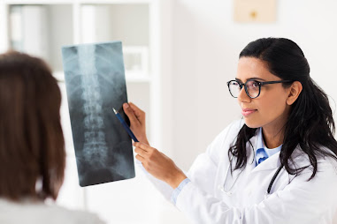
  What is Scoliosis? The spinal column should be straight when either viewed from the front or back. With scoliosis, there is a side shift of the spine to the right or left. Approximately 10% of the population has small curves (less than ten degrees), which are not any consequence to function or health. This condition is called Spinal Asymmetry. In the diagnostic x-ray, the curve of the spine measures in degrees, as an angle known as Cobb angle. Scoliosis is a curve greater than ten degrees. It occurs within the thoracic and lumbar regions of the spine and may involve one or both of those regions. The most common curve pattern may be a right thoracic curve. They occur within the vertebral column when viewed from the side (laterally). Round back (kyphosis) exists within the thoracic spine & Swayback (lordosis) occurs within the cervical and lumbar regions . What causes scoliosis? Scoliosis can arise from several underlying conditions, but is generally idiopathic, means “cause un...

MISS – Minimally Invasive Spine Surgery

Image
  Minimally Invasive Spine Surgery (MISS) may be a general term wont to describe a spread of surgical techniques which involve making smaller incisions and reducing the quantity of tissue damage beneath the skin. Some specially designed instruments assist in the visualization of the surgical field. It occurs with tubular retractors, allowing the surgeon to form a small incision but visualize an outsized area beneath the tiny incision.   Benefits of Minimally Invasive Spine Surgery:    Small scars following the operation causing lesser damage to the encompassing tissues beneath the skin.  Reduced pain, less blood loss Best cosmetic outcomes from smaller skin incisions Reduced muscle damage Reduced risk of infection and postoperative pain Faster recovery from surgery and low rehabilitation  Diminished reliance on pain medications after surgery MISS surgeries also can be performed under local anaesthesia and as daycare pr...

Recovery from a ruptured intervertebral disk

Image
  A ruptured disc causes severe low back pain and, sometimes, shooting pain down the rear of the legs, mentioned as sciatica. Usually, the symptoms of a disc rupture heal naturally by-themselves after a couple of weeks to a month. If the matter persists for months and becomes chronic, you will like better to consider surgery. Ruptured discs become increasingly usual with ageing and hence the breakdown of the spinal discs. It is difficult to prevent a ruptured disc, but regular back-strengthening exercise can lower your risk. Recovery Most disc pain substantially improves within a month. Expect gradual improvement after the first acute right stage after the flare-up. Exercise can help to stop the outburst of disc pain in future. Traditional exercise like yoga can help tone and strengthen the core muscles, supporting your spine. Be mindful that you don't overdo it with any exercise since that might trigger new back pain. Disc wear and tear tends to worsen over time, so you would like...

How do I find a good neurologist?

Image
  If it is time for you to see a neurologist, you have another vital decision to make—choosing the best neurologist who is apt for you or a family member. You will depend on your neurologist knowledge and expertise to diagnose neurological disease and reduce neurological disability. How do you find the best neurologist who is right for you? Here are some crucial factors to keep in mind while selecting a neurologist: 1. Get Referrals 2. Research the Neurologist Credentials 3. Consider the Neurologist Experience 4. Consider Gender 5. Research Hospital Quality  6. Evaluate Communication Style .7.  Read Patient Reviews 8. Know What Your Insurance Covers Your doctor's hospital is your hospital. For this reason, consider the quality of care at the hospital & get comfortable with your neurologists & the nursing staff at the hospital.   For more information for the best neurological diagnosis, treatment & care from the renowned neurologists Visit us: https://www....

What is Trigeminal Neuralgia (TN)?

Image
  Trigeminal neuralgia is a pain disorder that affects the fifth cranial nerve, the trigeminal nerve. This nerve controls the functions like chewing and biting and carries sensations from the face to the brain. Though commonly seen in people above 50 years of age, trigeminal neuralgia can occur at any age. Studies confirm that TN is more common in women than men. What are the causes of TN? An inflated artery or vein, injury, tumour, inflammation of meninges or other lesions may compress or irritate the trigeminal roots. It damages the protective coating around the nerve (myelin sheath), leading to the symptoms of trigeminal neuralgia. What are the signs and symptoms of TN? The patient experiences excruciating, intense, stabbing or shock-like pain over the eyes, lips, nose, forehead, mandible and upper jawbone. An attack may extend from a few seconds to several minutes and may occur several times a day. There might also be a persistent stabbing or aching pain in the area of the face...

Bulging Disc Vs. Herniated Disc: What's The Difference?

Image
What is a herniated disc? When a disc herniates, the soft nucleus spurts out through a tear within the annulus. Because there is limited space in the spinal canal, it can compress a nerve root or the spinal cord. The nucleus can extrude into the spinal canal, foramen, on either side of the disc or in rare cases, in multiple places called multifocal extruded discs. Typically, a disc that herniates is within the final stages of degeneration. Although herniated discs can occur anywhere in the spine, they are more common in the lumbar region. Herniated discs are also known as disc extrusion, slipped disc, or ruptured disc. What is a bulging disc? Unlike a herniated disc, when the nucleus breaks through the annulus, a bulging disc protrudes outward with the outer layers of the annulus remain intact. However, because the disc protrudes into the vertebral canal, it can still compress a nerve root. A disc bulge is also called disc prolapse. Types of Herniated and Bulging Discs Herniated and Bu...

Lumbar Disc Herniation-Symptoms, Causes, Treatment, Recovery and Exercises!!

Image
  Lumbar Disc Herniation-Symptoms, Causes, Treatment, Recovery and Exercises The vertebrae are the bony building blocks of the human spine. Between each of the parts of the vertebrae are the discs with ligaments situated around the spine and discs. The spinal has seven vertebrae within the neck (cervical vertebrae), 12 vertebrae within the mid-back (thoracic vertebrae), and five vertebrae within the low back (lumbar vertebrae). Within the mid-buttock, beneath the fifth vertebra, is that the sacrum, followed by the tailbone. The bony spine designed so that vertebrae “stacked” together can provide a movable support structure while also protecting the medulla spinals (nervous tissue that extends down the vertebral column from the brain) from injury. Each vertebra features a spinous process, a bony prominence behind the medulla spinals that shields the cord’s nervous tissue. The vertebrae even have a robust body ahead of the medulla spinals to supply a platform suitable for weight-bear...

What does a brain aneurysm look like?

Image
  A bulge or ballooning in a blood vessel in the brain is called a  brain aneur ysm .   A brain aneurysm occurs when a weak spot in the brain’s arterial wall bulges and fills with blood, they develop as a result of the thinning of artery walls and are commonly seen at the branches of the arteries which are the weakest points of the vessels. Although aneurysms can appear anywhere within the brain, they are most commonly found in arteries at the base of the brain. A brain aneurysm is a potentially life-threatening condition that may affect an individual at any age. If a brain aneurysm ruptures, it’s an emergency situation that may lead to a stroke, brain damage, and even death if not treated immediately. A ruptured which causes bleeding inside the brain is called hemorrhagic stroke, and the one occurring in the space between the brain and the thin tissues covering the brain is called a subarachnoid hemorrhage. Most brain aneurysms are often detected during tests for ot...
                       Awake Brain Surgery- A Fact and not a Fiction! What is Awake Brain Surgery (Intraoperative brain mapping)? Neurosurgeons perform many brain tumour procedures while the patient is awake but sedated called Awake Brain Surgery. An Awake brain surgery is to treat some brain (neurological) conditions, including some brain tumours or epileptic seizures. It enables the neurosurgeons to get rid of tumours that might rather be inoperable because: · They are on the brink of areas of the brain that control vision, language and body movements · The surgery may result in a loss of these functional areas. When the tumours have spread throughout the brain without borders (sorts of glioma), awake brain surgery is performed. An Awake brain surgery can shrink these tumours. During the surgery, your surgeon asks you questions and accordingly monitors the brain activity as yo...